
Authors:
F.J. Voskuil, MD, P.J. Steinkamp, MD, M.J.H. Witjes, MD, DDS, PhD & G.M. van Dam, MD, PhD
We believe that life for clinicians is getting easier when intuitive technology can help in difficult decision making during the medical evaluation and therapy process. Imagine, would you rather differentiate between white and beige, or between white and red? Indeed, the more discriminative the colors, the easier it will be. That is exactly the unmet need surgical oncologists are facing every day in clinical practice.
In surgical oncology, the ultimate goal of treatment is exciding all tumor tissue, while respecting the adjacent healthy tissue. A clear discrimination between ‘the good’ and ‘the bad’ is therefore essential. Unfortunately, to date, no clinical tool is available that can provide in this demand. For more than a decade, a still growing research field has been established that is convinced that lighting up tumors might be the solution to the desire of real-time intra-operative information. In 2011, Go van Dam and colleagues published for the first time about the clinical application of a fluorescent optical imaging agent targeting ovarian cancer in Nature Medicine1. From then on, dozens of clinical studies have been performed in a variety of cancer types. Our group, as well as others, have been investigating a broad range of solid tumors. In most studies, a tumor-type specific antibody, conjugated to a fluorescent dye was used for highlighting the cancerous tissue of interest by targeting a cell-surface receptor 2-6. Indeed, the potential of these strategies is undoubtedly very high, and actually some imaging agents are currently investigated in large clinical phase II/III pivotal studies. Despite the advancements towards clinical application, there are some limitations which might hamper broad clinical applicability of some of these antibody-based imaging agent strategies. Since cancer is a heterogenous disease, receptor expression might differ within and between patients. Moreover, as we know, even in ‘the good’ (i.e. healthy tissue), background target expression might be present, which can limit the discriminative strength of the imaging agent to detect cancer cells. Our colleagues and co-authors of the current paper – Jinming Gao and Baran Sumer – strongly believed that another more generic imaging strategy might be the next revolutionary step for optical imaging in surgical oncology. In contrast to targeting the diverse geno-/phenotypes of different types of cancer, they believed it was rationally logical and worth trying to target the deregulated energetics, considered to be a hallmark of cancer 7. Tumor acidosis is a general phenomenon observed in solid tumors already observed almost 100 years ago by the German physiologist and Nobel Laureate, Dr. Otto Warburg in 1924. Despite extensive research into this distinctive metabolic phenotype, tumor acidosis has never been utilized and proven to be clinical feasible as an imaging target for diagnostic or therapeutic purposes. The preclinical research results obtained by the group of Gao et al led to the GMP production of a pH-sensitive tracer and drug delivery compound, called ONM-100, which opened-up the initiation of a first-in-human clinical trial investigating the clinical applicability of tumor acidosis in cancer patients.
Clinical application of the drug
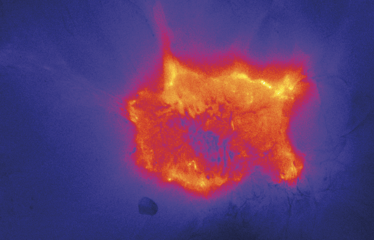
From the moment professor Go van Dam – surgeon oncologist and corresponding author on this paper – came in contact with this imaging agent and the possibility of joining forces to explore the clinical utility of ONM-100, he immediately had the feeling that this could be the next generation optical imaging agent for solid cancers. After a long journey of logistical, regulatory and scientific challenges, Floris Voskuil and Pieter Steinkamp – first authors on this paper - started the clinical trial in March 2018. The goal was to include thirty patients. Since ONM-100 had the potential to fluoresce in tumors irrespectively of the tumor-type, we decided to include four different solid tumor types (Head and Neck Squamous Cell Cancer, Breast Cancer, Esophageal Cancer and Colorectal Cancer). Back to that day in March: administering a new drug to a patient for the first time in the world - sounds awesome by the way - is exciting but also scary. Very scary by the way for any first in-human application of any compound, especially for two young medical doctors. Therefore, a solid back-up team was arranged and available, just in case… The intravenous administration of the drug went without any problem, and preparations were made for the surgery scheduled for the next day, a mastectomy (removal of the complete breast). The team was thrilled, excited and nervous at the same time with a lot of questions: Will it work? Do we see any fluorescence at all…..? Immediately after surgery the freshly excised specimen was cut in bread-loaf slices at the Department of Pathology and one slice at the time was imaged real-time. The first slices did not reveal any fluorescence (which might be a good sign, since no tumor was expected at the borders of the excision specimen). Then slice number 8 was imaged…… After processing the image, a loud shout was heard by the whole department. Floris and Pieter (and Go van Dam and Max Witjes - head and neck surgical oncologist and co-author on this paper - using FaceTime) observed fluorescence exactly at the position where the tumor was (Figure 1)! The feeling you get as a young scientist and the complete team of collaborators – also the ones we woke-up in the middle of the night in the USA - is amazing, unique and unforgettable when a revolutionary innovative imaging strategy on which so many people have worked works in a clinical setting.

Figure 1 | The first image obtained of ONM-100 in breast cancer
Fluorescence image of a tissue slice containing invasive breast cancer. The dark core of the lesion corresponds to fibrotic/necrotic tissue which contains no viable tumor tissue.
Tumor-agnostic characteristics and results in perspective
As mentioned, we included thirty patients with different solid tumor types in this study. The results showed that irrespective of the investigated tumor type, a solid consistent tumor-to-background ratio is observed in all patients, which clearly shows the tumor-agnostic characteristics of ONM-100. Moreover, no worrying study related adverse events were found up to day 17 after administration of the drug. Even of greater clinical relevance is the 100% sensitivity of the detection of tumor-positive surgical resection margin. Briefly, immediately after excision the resection specimen were imaged at all resection margins. In all cases a histopathological proven tumor-positive surgical resection margin was present, which was correctly identified using ONM-100 fluorescence upfront. The clinical potential of these results is that in the very near future the excision specimen can be scanned for fluorescence positive lesions, and if any, an immediate re-resection can be performed by the surgeon while the patient is still anesthetized. We hypothesize that post-operative treatment strategies correcting for failed initial surgery can therefore be reduced for the benefit of the individual patient. Moreover, in several patients included in our study additional tumor lesions were detected using ONM-100 both in situ as during histopathological processing of the tissue, which were otherwise missed by standard of care. The above clearly indicates that fluorescence imaging is a robust, reproducible and clinical feasible imaging tool to assist the clinician in clinical decision-making, whether it is the surgeon or pathologist in this case for the better of the patient(s) involved.
By writing this Behind the Paper, this trial has come to a successful end, but the journey towards clinical implementation of next generation imaging tools for enhancing cancer surgery will continue, with a bunch of nice colleagues and friends together at a global scale all for the cancer patients of today and tomorrow!
References:
- van Dam, G. M. et al. Intraoperative tumor-specific fluorescence imaging in ovarian cancer by folate receptor-alpha targeting: first in-human results. Nat Med 17, 1315–1319 (2011).
- Voskuil, F. J. et al. Fluorescence-guided imaging for resection margin evaluation in head and neck cancer patients using cetuximab-800CW: A quantitative dose-escalation study. Theranostics 10, 3994–4005 (2020).
- Rosenthal, E. L. et al. Safety and Tumor Specificity of Cetuximab-IRDye800 for Surgical Navigation in Head and Neck Cancer. Clin. Cancer Res. 21, 3658–3666 (2015).
- Gao, R. W. et al. Safety of panitumumab-IRDye800CW and cetuximab-IRDye800CW for fluorescence-guided surgical navigation in head and neck cancers. Theranostics 8, 2488–2495 (2018).
- Lamberts, L. E. et al. Tumor-Specific Uptake of Fluorescent Bevacizumab-IRDye800CW Microdosing in Patients with Primary Breast Cancer: A Phase I Feasibility Study. Clin. Cancer Res. (2016).
- Koller, M. et al. Implementation and benchmarking of a novel analytical framework to clinically evaluate tumor-specific fluorescent tracers. Nat Commun 9, 37–39 (2018).
- Zhao, T. et al. A transistor-like pH nanoprobe for tumour detection and image-guided surgery. Nat Biomed Eng 1, 1–8 (2016).


Please sign in or register for FREE
If you are a registered user on Research Communities by Springer Nature, please sign in