Study rationale
Breast cancer screening has been a phenomenal success in catching cancer at an earlier, more treatable, stage. However, with this comes the prospect of overtreating women with a precancerous form of breast cancer, ductal carcinoma in situ (DCIS), that may never progress to invasive disease. Currently there is no way to differentiate women whose DCIS will progress from those whose disease will remain indolent.
With a lack of distinct genetic changes between the cancer cells in DCIS and invasive disease, it is suggested that the most critical changes underpinning DCIS progression lie in the surrounding microenvironment1. Understanding the role of the tumour microenvironment will shed light on the critical steps that drive the transition of DCIS to invasive cancer and identify candidate biomarkers, which could be used to stratify high-risk patients and reduce unnecessary treatment.
What did we show?
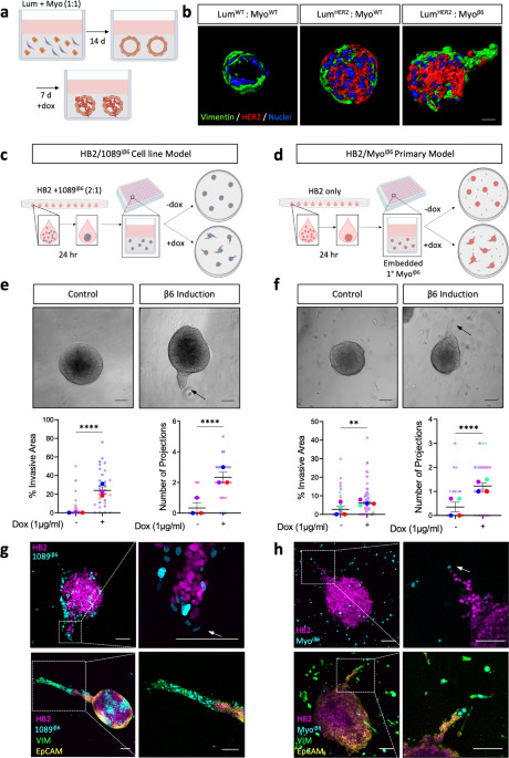
To understand the mechanisms that drive progression of DCIS to invasive cancer, we developed several 3D spheroid models of the human breast duct, incorporating different cell types of the breast into a physiologically relevant matrix. We show that myoepithelial cells, a non-cancerous cell-type in the microenvironment, can adopt a tumour-promoting phenotype upon induced upregulation of integrin β6, a previously established marker of high-risk DCIS2. Using cell-type specific fluorescent labels, we observe that β6-expressing myoepithelial cells drive the invasion of pre-neoplastic luminal cells into the surrounding matrix.
Subsequent transcriptomic analysis revealed changes in the expression of proteolytic enzymes called MMPs, that degrade surrounding matrix proteins. We show that β6-mediated myoepithelial-led invasion is dependent on myoepithelial MMP13 and can be blocked by targeting MMP13 using genetic or pharmacological inhibition. Analysis of downstream signalling pathways shows that β6-mediated MMP13 upregulation is dependent on TGFβ signalling and subsequent activation of the histone acetyltransferase EP300. Interestingly, interrogation of publicly available data sets shows that high expression of EP300 in breast cancer patients is associated with poorer prognosis. Importantly, we confirm our 3D model findings in clinical tissue and demonstrate that MMP13 expression is observed in the myoepithelial cells of DCIS, but not normal, healthy ducts3.

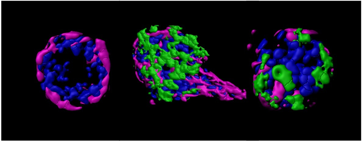
Figure 1. (A) Fluorescent images of invading heterocellular HB2/1089iβ6 spheroids upon integrin β6 induction with HB2 luminal (magenta) and 1089iβ6 myoepithelial (cyan) cells. (B) Light micrographs of heterocellular HB2/1089iβ6 spheroids following integrin β6 induction and/or myoepithelial siRNA-mediated knockdown of MMP13. (C) We propose a mechanism whereby myoepithelial integrin β6 upregulation in DCIS promotes TGFβ/EP300-mediated upregulation of MMP13 to facilitate invasive progression. Scale bar = 100 µm.
What is surprising about our results?
Myoepithelial cells are natural tumour suppressors of the healthy breast duct, where they act as a physical barrier to stop invading luminal cells from escaping to the surrounding breast tissue. In our study, we show that during the initial stages of breast cancer, these myoepithelial cells can become dysfunctional and lose their tumour suppressive function. They start to drive invasion of luminal cells and facilitate the progression of DCIS to invasive cancer, turning the notion that myoepithelial cells are exclusively tumour suppressive on its head.
What are the implications of our study?
This study demonstrates the power of 3D model systems as a discovery tool leading to new mechanistic insights and clinically relevant discoveries. 3D models of the tumour microenvironment allow complex cellular interplay to be dissected in exquisite detail within physiologically relevant contexts4. Their increased use will reveal new biology indiscernible in traditional 2D systems, reducing the need for animal models.
We hope to validate myoepithelial MMP13 expression in a larger patient cohort with long-term follow up data, where the endpoint assessed is the appearance of any recurrent disease. This will allow us to assess the predictive value of myoepithelial MMP13 as an appropriate biomarker for high-risk DCIS patients.
References
- Gibson SV, Roozitalab RM, Allen MD, Jones JL, Carter EP, Grose RP. Everybody needs good neighbours: the progressive DCIS microenvironment. Trends in Cancer. S2405–8033, 00002-X. (2023) Epub ahead of print.
- Allen MD, Thomas GJ, Clark S, Dawoud MM, Vallath S, Payne SJ, et al. Altered microenvironment promotes progression of preinvasive breast cancer: myoepithelial expression of alphavbeta6 integrin in DCIS identifies high-risk patients and predicts recurrence. Clin Cancer Res. 2014;20(2):344-57.
- Gibson SV, Tomas Bort E, Rodríguez-Fernández L, Allen MD, Gomm JJ, Goulding I, et al. TGFβ-mediated MMP13 secretion drives myoepithelial cell dependent breast cancer progression. npj Breast Cancer. 2023;9(1):9.
- Carter EP, Roozitalab R, Gibson SV, Grose RP. Tumour microenvironment 3D-modelling: Simplicity to complexity and back again. Trends in Cancer. 2021;7(11):1033-46.


Please sign in or register for FREE
If you are a registered user on Research Communities by Springer Nature, please sign in